Cryogenic ion spectroscopy is a cutting-edge technique used in combination with mass spectrometry for identification and structural characterization of small to mid-size biomolecules with unparalleled confidence and sensitivity.
How It Works
Step 1
Ionization
Biomolecules are first converted into ions, and introduced into a mass spectrometer.
Step 2
Cooling
The ions of interest are then cooled to cryogenic temperatures using a compact cryocooler. Cooling significantly reduces thermal broadening, leading to high-resolution spectra.
Step 3
Spectroscopy
Once cooled, the ions are fragmented by infrared or ultraviolet laser pulses at variable wavelength.
Step 4
Mass Spectrometry
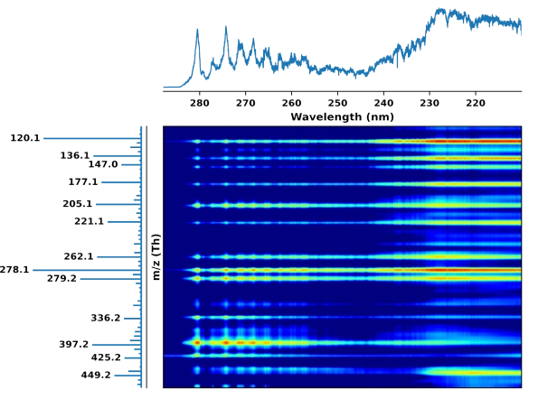
At each wavelength the fragment ions are monitored by high resolution MS. The recorded 2D UV/IR-MS fingerprint uniquely reflects structure of the ions.
Applications
Unambiguous identification and fast quantification of isomers
Our 2D UV-MS fingerprinting method enables library-based and library-free identification and quantification of isomers in seconds
Non-targeted analysis of metabolites
Cryogenic ion spectroscopy as an orthogonal dimension for metabolite analysis
Intrinsic 3D structure of biomolecules
Combining cryogenic infrared ion spectroscopy with quantum-chemical calculation allows for accurate elucidation of 3D molecular structure
Benefits
Structural Insights
This technique provides a unique look at the structure of biomolecules, helping scientists understand their functions and interactions
Versatile Applications
Cryogenic ion spectroscopy is highly versatile technology, applicable for identification and quantification of wide range of biomolecules and their isomers
Fast
Library-based identification and quantification can be done in seconds
High Sensitivity
Our technology works at MS level of sensitivity what is crucial for many life science applications
Compatibility
Cryogenic ion spectroscopy is compatible with LC-MS workflow